
Section 5 · Diagnostics and Preoperative Planning
Comprehensive trichological assessment of the surgical candidate: from history to the decision to operate
1.Clinical abstract and executive summary
Most hair transplant candidates have androgenetic alopecia, a condition whose prevalence in men rises progressively with age1. The outcome of a hair transplant is largely decided before surgery. Harvesting and implantation technique matter, but a wrong indication, an overestimated donor area or unforeseen progression of hair loss compromise even the best execution. The trichological assessment of the surgical candidate therefore has four aims: to confirm the diagnosis, to estimate future progression, to measure donor capacity and to check that the patient’s expectations are compatible with what surgery can offer.
This pillar article describes a structured six-step pathway: history, clinical examination, trichoscopy, measurement of the donor area and hair characteristics, photographic documentation, and assessment of expectations and psychological status. Each step is linked to the surgical decisions that follow from it. Specific aspects, such as calculating the safe donor area and designing the hairline, are covered in the other articles of section 5.
- The diagnosis must be confirmed even when it seems obvious: androgenetic alopecia can coexist with an effluvium or a cicatricial alopecia4,5.
- Planning is based on the expected progression of hair loss, not on the situation on the day of the visit2.
- The donor area is a finite resource: it must be measured, not estimated by eye.
- Unrealistic expectations and body dysmorphic disorder are relative contraindications that must be recognised before operating10.
2.Microscopic and ultrastructural anatomy: what is measured
Assessment of the candidate translates into numbers the anatomy described in the articles of section 1. The units of measurement are the follicular unit and the individual hair.
- Follicular unit density. The number of units per square centimetre in the donor area. In the Caucasian scalp it generally ranges from 65 to 100 units/cm² and decreases with age.
- Hairs per unit. The mean number of hairs in each unit, usually slightly more than 2. Together with unit density, it determines hair density.
- Shaft calibre. A larger-diameter hair covers more: for the same number of grafts, a high calibre produces a greater impression of density.
- Morphology and colour. Wavy or curly hair covers more surface than straight hair; low contrast between hair colour and skin colour makes thinning less visible.
- Follicular depth and angle. These vary by region and influence the harvesting technique9.
These parameters describe not only the quantity of hair available but also its capacity to cover a surface. Two patients with the same number of available grafts can achieve very different visual results.
3.Molecular dynamics and signalling pathways: the role of biomarkers
There is currently no validated molecular biomarker that predicts, in the individual patient, the rate of progression of androgenetic alopecia or the stability of the donor area. Genome-wide studies have identified dozens of loci associated with male pattern baldness11, but their individual predictive value remains limited and commercial genetic tests currently have no role in surgical planning.
Assessment therefore remains clinical. Its “markers” are indirect: family history, age at onset, the stage reached at a young age and, above all, the degree of miniaturisation documented on trichoscopy, which reflects the effect of androgens on the dermal papilla of each region. Miniaturisation in the recipient area identifies the hairs at risk; its presence in the donor area signals a donor that is not fully stable.
4.Pathophysiology and clinical correlations: the six-step pathway
1. History
The trichological history records age at onset, perceived rate of progression, family history on both sides, current or previous treatments and their response, and any previous surgery. Events in the three months before the visit, such as fever, surgery, drugs, diets and stress, should be explored, as they may point to a superimposed telogen effluvium4. In women, the menstrual cycle, signs of hyperandrogenism, pregnancies and hormone therapy are relevant. Systemic conditions, anticoagulant and antiplatelet drugs, allergies and a tendency to hypertrophic scars or keloids complete the picture.
2. Clinical examination
Physical examination classifies the pattern of hair loss: the Hamilton-Norwood scale in men2 and the Ludwig scale in women3. The pull test checks whether active shedding is under way. The scalp skin, eyebrows and body hair should be examined to rule out signs of frontal fibrosing alopecia or alopecia areata5. Finally, the laxity of the donor area is assessed, which helps in choosing between FUE and FUT.
3. Trichoscopy
Trichoscopy is the core of the assessment6. It should be performed at a minimum of three sites: frontal region, vertex and occipital area. Comparing recipient and donor areas makes it possible to quantify anisotrichosis, that is, variability in shaft diameter greater than 20%, and the proportion of miniaturised hairs7. Trichoscopy also identifies signs of alopecias other than androgenetic alopecia: loss of follicular openings, perifollicular erythema and scaling, yellow dots and exclamation-mark hairs.
4. Donor area measurements
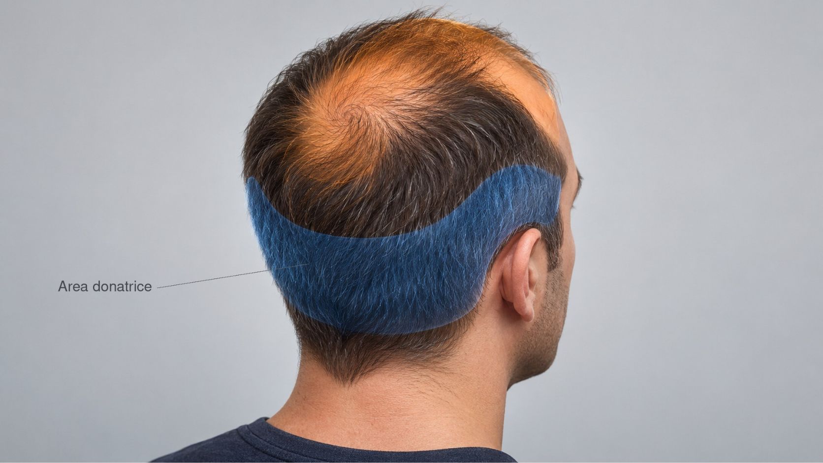
In the donor area, follicular unit density, the mean number of hairs per unit and calibre are measured at several sites, because values vary between the occipital, parietal and temporal regions. The estimate of grafts available over a lifetime is derived from the surface of the safe donor area, the density and the proportion of units that can be harvested without producing visible thinning. The calculation method is described in the articles on the safe donor area and the follicular budget.
5. Photographic documentation
Standardised photographs, with the same views, the same distance, the same lighting and the hair in the same condition, are essential for three reasons: they document the starting point, they allow disease progression to be distinguished from the effect of surgery, and they protect both doctor and patient in the event of a dispute.
6. Expectations and psychological status
The consultation must clarify what the patient expects and whether this expectation can be achieved with the available resources. Body dysmorphic disorder, which is more common among people seeking aesthetic treatments than in the general population, must be considered: disproportionate concern about a minimal or invisible defect calls for specialist assessment before any procedure10.
| Step | Key elements | Decision influenced |
|---|---|---|
| History | Onset, family history, treatments, recent events, drugs | Diagnosis, timing, medical preparation |
| Clinical examination | Classification, pull test, eyebrows, laxity | Differential diagnosis, choice of FUE or FUT |
| Trichoscopy | Miniaturisation at three sites, signs of other alopecias | Indication, donor stability, risk of shock loss |
| Donor measurements | Unit density, hairs per unit, calibre | Number of grafts available, number of sessions |
| Photographs | Standardised views and lighting | Comparison over time, medico-legal protection |
| Consultation | Expectations, body dysmorphia | Indication, informed consent |
5.Surgical protocols and impact on FUE microsurgery
Every piece of information gathered during the assessment translates into an operative choice.
- Choice of technique. Laxity, density and hair characteristics guide the choice between FUE and FUT. FUE is preferable when laxity is low or the patient wears short hair; FUT may be considered when many grafts are needed in a single session and laxity allows it.
- Punch diameter and depth. Hair morphology and curvature, observed on trichoscopy and with test extractions, guide the choice of punch diameter and incision depth9.
- Graft distribution. The number of grafts available over a lifetime sets the priorities: in general the frontal region and hairline come before the vertex, because they have the greatest impact on facial appearance.
- Implantation density. The proportion of miniaturised native hairs in the recipient area determines implantation density and the risk of shock loss.
- Number of sessions. When the need exceeds what can safely be harvested at once, planning includes several spaced sessions.
6.Regenerative medicine and adjuvant treatments
The assessment also defines the role of medical therapy. In candidates with progressing androgenetic alopecia, the European S3 guideline identifies oral finasteride and topical minoxidil as the treatments with the highest level of evidence8; the full text is available on the European Dermatology Forum website. Starting therapy a few months before surgery makes it possible to assess the response, stabilise native hair and, in some cases, reduce the number of grafts needed. Platelet-rich plasma and other regenerative treatments may be considered as an adjunct, bearing in mind that the evidence is of moderate quality and protocols are not standardised.
7.Risk management and anatomical limits
- Young patients. Before the age of 25–30 the course of hair loss is hard to predict: planning must be cautious and sustainable even in a scenario of advanced progression.
- Limited donor area. Low density, fine hair or miniaturisation in the occiput reduce surgical possibilities and must be communicated clearly.
- Unconfirmed diagnosis. Signs of cicatricial alopecia, alopecia areata or ongoing effluvium require surgery to be postponed or ruled out5.
- Systemic conditions and drugs. Coagulopathies, anticoagulant therapy and uncontrolled disease require dedicated preoperative medical assessment.
- Expectations. A request for density or a hairline that cannot be sustained by the available donor area should be discussed openly; if disagreement persists, it is better not to operate.
8.Clinical tips & tricks from the medical team
- An unhurried first visit. A complete assessment takes time. The decision to operate should not be made at the same visit at which the first history is taken.
- Measure at several sites. Donor density varies between the occiput, parietal regions and temples: a single measurement may overestimate overall capacity.
- Show the patient the trichoscopy. Seeing miniaturised hairs on the screen helps the patient understand why medical therapy and long-term planning are needed.
- Write down the plan. A written plan with the estimated number of grafts, areas treated, priorities and possible future sessions makes consent better informed and reduces misunderstandings.
- Reassess after therapy. In patients starting medical therapy, a new assessment after a few months makes it possible to recalibrate the surgical plan.
— Methodological approach of the Clinica Pallaoro team, Padua
9.Critical discussion and conclusions
Candidate assessment is the least studied phase of hair restoration surgery. Most selection criteria derive from clinical experience and expert consensus rather than from prospective studies. There are no validated tools for predicting individual progression, and donor area measurements vary with the method and the examiner’s experience. Tools such as the phototrichogram, digital image analysis and, in the future, artificial intelligence systems could make these measurements more reproducible.
In the meantime, a structured assessment remains the best tool available. The surgeon who confirms the diagnosis, measures the donor area, documents the starting point and checks the patient’s expectations makes more cautious decisions that are more sustainable over time. The decision not to operate, when conditions do not allow it, is fully part of good surgical practice.
10.Annotated scientific bibliography
- Severi G, Sinclair R, Hopper JL, et al. Androgenetic alopecia in men aged 40-69 years: prevalence and risk factors. Br J Dermatol. 2003;149(6):1207-1213. University of MelbournePrevalence and risk factors of androgenetic alopecia in adult men.
- Norwood OT. Male pattern baldness: classification and incidence. South Med J. 1975;68(11):1359-1365. PubMedThe Hamilton-Norwood classification and its distribution by age.
- Ludwig E. Classification of the types of androgenetic alopecia (common baldness) occurring in the female sex. Br J Dermatol. 1977;97(3):247-254. PubMedReference classification of female androgenetic alopecia.
- Headington JT. Telogen effluvium. New concepts and review. Arch Dermatol. 1993;129(3):356-363. PubMedFunctional classification of telogen effluvium and the interval between trigger and shedding.
- Kossard S. Postmenopausal frontal fibrosing alopecia. Scarring alopecia in a pattern distribution. Arch Dermatol. 1994;130(6):770-774. PubMedFrontal fibrosing alopecia: differential diagnosis of frontal recession.
- Rudnicka L, Olszewska M, Rakowska A, Kowalska-Oledzka E, Slowinska M. Trichoscopy: a new method for diagnosing hair loss. J Drugs Dermatol. 2008;7(7):651-654. PubMedTrichoscopy as a diagnostic method for alopecia.
- Rakowska A, Slowinska M, Kowalska-Oledzka E, Olszewska M, Rudnicka L. Dermoscopy in female androgenic alopecia: method standardization and diagnostic criteria. Int J Trichology. 2009;1(2):123-130. PMCStandardised trichoscopic criteria, including the anisotrichosis threshold.
- Kanti V, Messenger A, Dobos G, et al. Evidence-based (S3) guideline for the treatment of androgenetic alopecia in women and in men – short version. J Eur Acad Dermatol Venereol. 2018;32(1):11-22. PubMedEuropean guideline for the medical treatment of androgenetic alopecia.
- Jiménez F, Izeta A, Poblet E. Morphometric analysis of the human scalp hair follicle: practical implications for the hair transplant surgeon and hair regeneration studies. Dermatol Surg. 2011;37(1):58-64. University of MurciaMorphometric data on the scalp follicle with implications for the surgeon.
- Phillips KA, Dufresne RG Jr. Body dysmorphic disorder. A guide for dermatologists and cosmetic surgeons. Am J Clin Dermatol. 2000;1(4):235-243. Weill Cornell MedicineRecognition and management of body dysmorphia in patients seeking aesthetic treatments.
- Heilmann-Heimbach S, Herold C, Hochfeld LM, et al. Meta-analysis identifies novel risk loci and yields systematic insights into the biology of male-pattern baldness. Nat Commun. 2017;8:14694. PMCGenome-wide meta-analysis of genetic loci associated with male pattern baldness.
Content intended for healthcare professionals for continuing scientific education. It does not replace individual clinical assessment. © Clinica Pallaoro — trapiantocapelli.com